Home
/ Back Of Neck Nerves Anatomy - Common Back And Neck Conditions Boston Orthopaedic Spine : They innervate the posterior scalp up as far as the vertex and other structures as well, such as the ear.
Back Of Neck Nerves Anatomy - Common Back And Neck Conditions Boston Orthopaedic Spine : They innervate the posterior scalp up as far as the vertex and other structures as well, such as the ear.
Back Of Neck Nerves Anatomy - Common Back And Neck Conditions Boston Orthopaedic Spine : They innervate the posterior scalp up as far as the vertex and other structures as well, such as the ear.. The seven small vertebrae that begin at the base of the skull and form the neck comprise the cervical spine. Nerves the accessory nerve (cn xi) exits the cranial cavity, descends down the neck, innervates sternocleidomastoid and enters the posterior triangle. It crosses the posterior triangle in an oblique, inferoposterior direction, within the investing layer of fascia. In addition, in this region we also find the major cranial and spinal nerves that connect the central nervous system to the organs, skin, and muscles of the head and neck. As they join, they form the spinal nerves on the sides of the spinal cord.
Peripheral nerves comprise the peripheral nervous system. A mixed nerve containing both motor and sensory fibers. Sympathetic innervation to the skin. The main arteries supplying the oral cavity are the descending palatine, facial, lingual, and maxillary arteries. These muscles support the spine and allow for movement.
Neck Pain Chiropractor Blog Back Pain And Headache Specialist Burke Va Nova Headache Chiropractic Center from images.squarespace-cdn.com Each nerve provides sensation to a specific area of the body called a dermatome. These muscles support the spine and allow for movement. Medical imaging studies may be conducted to determine the cause of nerve issues in the neck. These vertebrae have eight pairs of nerve roots located between the vertebrae, with nerve root pair c8 resting between the last cervical vertebra (c7) and the first thoracic vertebra (t1). The cervical column is comprised of 7 bones (c1 to c7) uniquely shaped to protect the spinal cord that descend from the base of your skull and the spinal nerves or root that exit the spine between each set of bones. A nerve that exits the lower back has peripheral branches that extend all the way down to the toes. These nerve roots emerge directly from the spinal cord—sensory nerve roots from the back of the spinal cord and the motor nerve roots from the front of the spinal cord. These 2 nerve roots branch directly from the spinal cord and merge to form the spinal nerve as it runs through an opening between adjacent vertebrae, called the intervertebral foramen.
The majority of these nerves control the functions of the upper extremities and allow you to feel your arms, shoulder, and back of your head.
The spinal nerves are relatively large nerves that are formed by the merging of a sensory nerve root and a motor nerve root. Hymen structure anatomy, function, types of hymen, hymen breakage, virginity relation. Contain the common carotid artery, internal. These muscles support the spine and allow for movement. The cervical spine has seven vertebrae, labeled c1 to c7, that start at the base of the skull and run through the neck and upper back. The spinal cord originates as a continuation of the brainstem, specifically the medulla oblongata, and travels within the vertebral canal.at the level of the l2 vertebra, the spinal cord becomes known as the cauda equina. The back functions are many, such as to house and protect the spinal cord, hold the body and head upright, and adjust the movements of the upper and lower limbs. In this page, we're going to study each of the above structures together with their respective branches. Peripheral nerves comprise the peripheral nervous system. There are three major occipital nerves in the human body: The cervical spine is the top part of the spine. C1, c2, and c3 (the first three cervical nerves) help control the head and neck, including movements forward, backward, and to the sides. It runs from the neck to the upper back.
It crosses the posterior triangle in an oblique, inferoposterior direction, within the investing layer of fascia. General sense (touch, pressure, pain, heat, cold, etc.) to the skin of the back. It runs from the neck to the upper back. The main arteries supplying the oral cavity are the descending palatine, facial, lingual, and maxillary arteries. The occipital nerves are a group of nerves that arise from the c2 and c3 spinal nerves.
Dorsal Trunk And Neck Neurovasculature Preview Human Anatomy Kenhub Youtube from i.ytimg.com More specifically, one end of the loop, the superior root, is derived from c1 (and possibly c2, depending on the literature), while the other, the inferior root, comes from c2 and c3. The content of the neck is grouped into 4 neck spaces, called the compartments. Nerves nerve roots exit the spinal cord in the neck and provide control and sensation to different parts of the body based on the spinal level from where they exit, including signals for movement of the head, neck, arms and fingers, breathing, and skin sensation in the upper body. It runs from the neck to the upper back. Tendons attach the muscles to the vertebrae. The occipital nerves are a group of nerves that arise from the c2 and c3 spinal nerves. Spinal anatomy is a remarkable combination of strong bones, flexible ligaments and tendons, large muscles and highly sensitive nerves. For more anatomy content please follow us.
These nerve roots emerge directly from the spinal cord—sensory nerve roots from the back of the spinal cord and the motor nerve roots from the front of the spinal cord.
These vertebrae have eight pairs of nerve roots located between the vertebrae, with nerve root pair c8 resting between the last cervical vertebra (c7) and the first thoracic vertebra (t1). Specifically, nerve c5 controls the deltoids and biceps. These nerve roots emerge directly from the spinal cord—sensory nerve roots from the back of the spinal cord and the motor nerve roots from the front of the spinal cord. The back's muscles start at the top of the back (named the cervical vertebrae) and go to the tailbone (also named the coccyx). Neck anatomy nerves picture there are 8 spinal nerves that originate from the cervical spine. The spinal cord originates as a continuation of the brainstem, specifically the medulla oblongata, and travels within the vertebral canal.at the level of the l2 vertebra, the spinal cord becomes known as the cauda equina. The cervical spine protects the nerves connecting to. Tendons attach the muscles to the vertebrae. The cauda equina is formed from the spinal nerves which arise from the end of the spinal cord. The spinal cord is a bundle of nerves that extends from the brain and runs through the cervical spine and thoracic spine (upper and middle back) prior to ending just before the lumbar spine (lower back). Some of the muscle groups the brachial plexus controls include the deltoids, biceps and pectoral muscles. Sympathetic innervation to the skin. The posterior root, located in back, carries sensory signals from the body back to the brain.
Compression of these nerves in the back can cause cauda equina syndrome, which. Medical imaging studies may be conducted to determine the cause of nerve issues in the neck. This section on the nerves of the neck discusses the anatomy of the cervical plexus and the phrenic nerves. Sympathetic innervation to the skin. These 2 nerve roots branch directly from the spinal cord and merge to form the spinal nerve as it runs through an opening between adjacent vertebrae, called the intervertebral foramen.
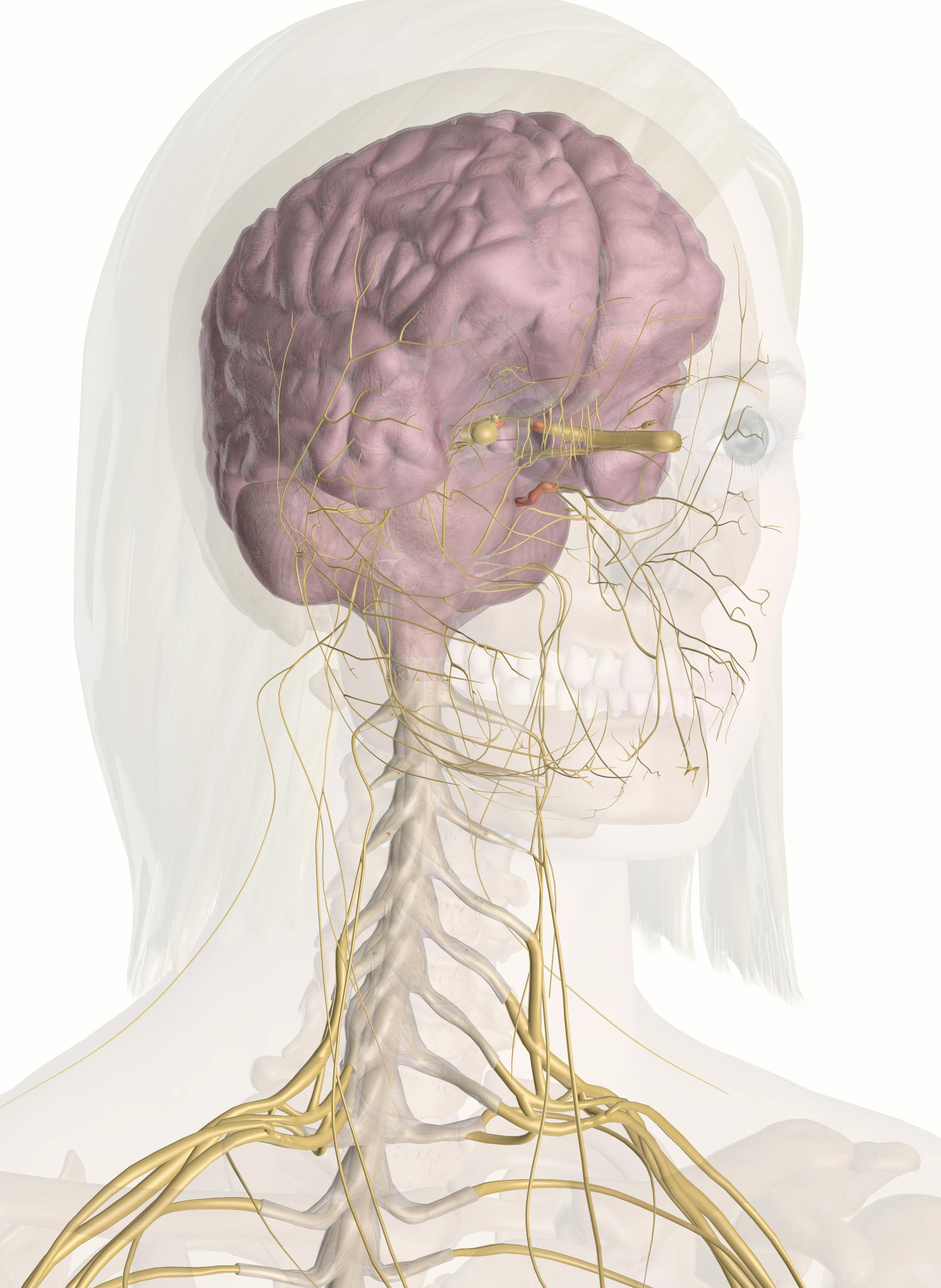
Nerves Of The Head And Neck Interactive Anatomy Guide from innerbody.imgix.net It comprises the vertebral column (spine) and two compartments of back muscles; The posterior root, located in back, carries sensory signals from the body back to the brain. These 2 nerve roots branch directly from the spinal cord and merge to form the spinal nerve as it runs through an opening between adjacent vertebrae, called the intervertebral foramen. Contains cervical vertebrae and postural muscles. This section on the nerves of the neck discusses the anatomy of the cervical plexus and the phrenic nerves. The majority of these nerves control the functions of the upper extremities and allow you to feel your arms, shoulder, and back of your head. In addition, in this region we also find the major cranial and spinal nerves that connect the central nervous system to the organs, skin, and muscles of the head and neck. For more anatomy content please follow us.
It comprises the vertebral column (spine) and two compartments of back muscles;
The ansa cervicalis (handle of the neck in latin), is a loop of nerves that lies superficial to the internal jugular vein, composed of the c1 to c3 nerves. Tendons attach the muscles to the vertebrae. General sense (touch, pressure, pain, heat, cold, etc.) to the skin of the back. Superior thyroid, ascending pharyngeal, lingual, facial, occipital, posterior auricular, maxillary, and superficial temporal arteries. The nerves of the head and neck include the most vital and important organs of the nervous system — the brain and spinal cord — as well as the organs of the special senses. Other parts of your spine include: A mixed nerve containing both motor and sensory fibers. The occipital nerves are a group of nerves that arise from the c2 and c3 spinal nerves. Hymen structure anatomy, function, types of hymen, hymen breakage, virginity relation. We hope this picture neck nerves and innervation of shoulder, arm, and hand can help you study and research. The cervical spine protects the nerves connecting to. In addition, in this region we also find the major cranial and spinal nerves that connect the central nervous system to the organs, skin, and muscles of the head and neck. Medical imaging studies may be conducted to determine the cause of nerve issues in the neck.
Contains glands ( thyroid, parathyroid, and thymus ), the larynx, pharynx and trachea back of neck anatomy. Some of the muscle groups the brachial plexus controls include the deltoids, biceps and pectoral muscles.

